
Presentation
Knee pain. A similar complaint was reported 9 months ago. The patient did not turn up for follow-up.
Patient Data
Age: 35 years
Gender: Male

Download
Info

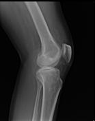
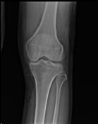
Knee radiograph - Serpentine channels seen in the suprapatellar region and Hoffa's region.


Download
Info
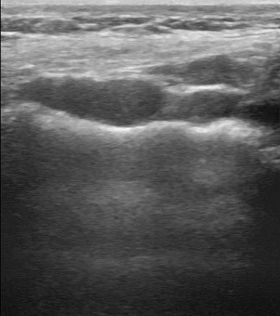
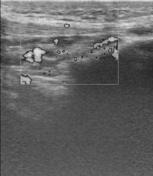
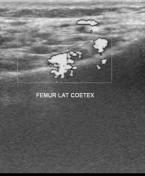
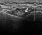
Multiple venous channels about knee - intra-articular as well as extra-articular.
First two images show femoral cortical erosion by the dilated veins.
Download
Info

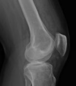
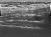
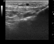
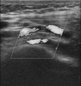
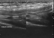
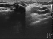
Dilated venous channels are noted deep to patellar and quadriceps tendons.
Case Discussion
Features of intra- and extra-articular venous malformation of the knee joint.


 Unable to process the form. Check for errors and try again.