
Presentation
Severe headache, no vomitting. Papilloedema
Patient Data
Note: This case has been tagged as "legacy" as it no longer meets image preparation and/or other case publication guidelines.
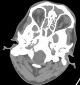

Basal sections through orbit show tortuous optic nerves on both sides.
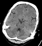
CT at basal ganglia level shows no supratentorial abnormality.
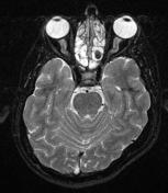

Axial fat sat T2WI through optic nerves shows optic nerve tortuosity.
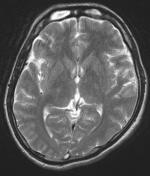
Axial T2WI shows loss of flow void in superior sagittal sinus.
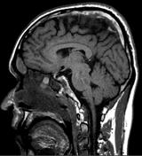
Sagittal T1WI shows normal sella and pituitary gland. Thrombus in superior sagittal sinus is noted.
Case Discussion
A patient presented for neuroimaging with h/o headache and decreasing vision for eight days. Clinical examination revealed bilateral papilloedema.
CT revealed normal neuroparenchyma with pansinusitis. Basal sections through the orbit revealed tortuous optic nerves on both sides.
MRI of brain and orbit done subsequently confirmed the findings seen on CT. In addition, MRI revealed extensive thrombosis of superior sagittal sinus and the right transverse sinus.
Lumbar puncture was performed; the CSF pressure was high (24cm), 30cc of CSF was let out and the patient was heparinised. The patient clinically improved with this therapy.


 Unable to process the form. Check for errors and try again.