
Presentation
Headache and blurring of vision. Papilloedema on clinical exam.
Patient Data


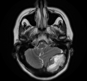
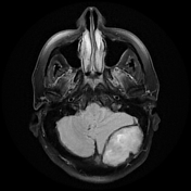
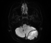

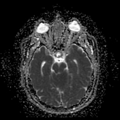
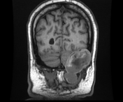
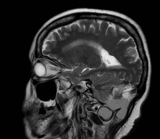
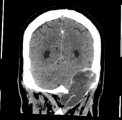
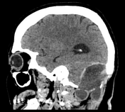
Left occipital intradiploic lesion eliciting mixed low/high signal on T1 and high signal on T2WI with diffusion restriction. It causes thinning of the overlying cortex. It displaces the left cerebellar hemisphere anteriorly with mild cerebellar tonsillar herniation, crowdening of the foramen magnum and subsequent mild hydrocephalic changes and signs of increased intracranial hypertension (empty sella and prominent perioptic CSF). It shows inferior extra-calvarial extension.
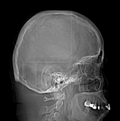
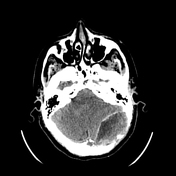
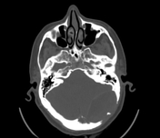

On CT scan, it appears lytic lesion with overlying cortical thinning.
Case Discussion
Intradiploic epidermoid cysts of the calvarium are very rare. They are derived from the ectodermal cells of the cranium and are lined solely by stratified squamous epithelium.
On CT, it appears lytic with scalloping and thinning of the outer and inner tables. On MRI, It elicits heterogeneous high signal on T1 and heterogeneous high signal on T2 WI with diffusion restriction and no/mild marginal post-contrast enhancement.


 Unable to process the form. Check for errors and try again.