Presentation
Painless swelling of the posterior aspect of the right upper leg.
Patient Data
![](https://prod-images-static.radiopaedia.org/images/61058977/5c75683f8ad7f175586ca04664873204b8cfc4d69e623581efbe7743993706c9_thumb.jpeg)
![This study is a stack](/packs/stack-YQKLCKBI.gif)
![](https://prod-images-static.radiopaedia.org/images/61058913/83d296b3a956478307347a53924dffbf437d8bb0b74006d80cff0f7cd934263d_thumb.jpeg)
![This study is a stack](/packs/stack-YQKLCKBI.gif)
![](https://prod-images-static.radiopaedia.org/images/61058930/045e6f6eec69f4edc1e1b85152cd9e16c3abf45d6915d358260e9e4242b411e9_thumb.jpeg)
![This study is a stack](/packs/stack-YQKLCKBI.gif)
![](https://prod-images-static.radiopaedia.org/images/61058953/d53060cdb76a8b8264320fc440408c936f1d85caa8d7d4b028fa6640080abf85_thumb.jpeg)
![This study is a stack](/packs/stack-YQKLCKBI.gif)
![](https://prod-images-static.radiopaedia.org/images/61059001/81c5afdb1e34fc2f05e13220907d491f766a80c956cada247576286b902a09f0_thumb.jpeg)
![This study is a stack](/packs/stack-YQKLCKBI.gif)
![](https://prod-images-static.radiopaedia.org/images/61059025/4d04f1c5383c6859a8c9f5c3c80beb952a97a92a12a494710390bb7c770bcf2e_thumb.jpeg)
![This study is a stack](/packs/stack-YQKLCKBI.gif)
![](https://prod-images-static.radiopaedia.org/images/61059073/dae7cfb255ea61e947f0db8251c652c83594be1b7287df29a0e0edcf6403d82d_thumb.jpeg)
![This study is a stack](/packs/stack-YQKLCKBI.gif)
![](https://prod-images-static.radiopaedia.org/images/61059193/4767cdfea4e6ed86cd859e5161e211f776e2fa04dd09623edf8629a4ffbc15b0_thumb.jpeg)
![This study is a stack](/packs/stack-YQKLCKBI.gif)
![](https://prod-images-static.radiopaedia.org/images/61059317/813958067bbb4eddfb10187071228f9014cb2d597ea385188ba3c10323ce5a83_thumb.jpeg)
![This study is a stack](/packs/stack-YQKLCKBI.gif)
![](https://prod-images-static.radiopaedia.org/images/61059339/77a2fcde86a86f52a306b3c3f5321956d55fc57dc7e74b4935d8bdf88958d6c0_thumb.jpeg)
![This study is a stack](/packs/stack-YQKLCKBI.gif)
![](https://prod-images-static.radiopaedia.org/images/61059362/70cd6fa0d936117fb5c26c2d156233b72cc8c96ecd924e19e89e2c259435a910_thumb.jpeg)
![This study is a stack](/packs/stack-YQKLCKBI.gif)
![](https://prod-images-static.radiopaedia.org/images/61058977/5c75683f8ad7f175586ca04664873204b8cfc4d69e623581efbe7743993706c9_big_gallery.jpeg)
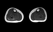
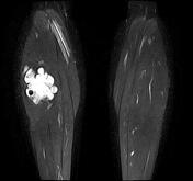
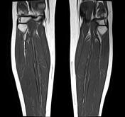
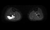
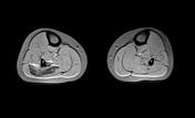
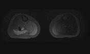
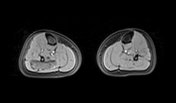
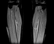
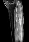
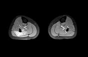
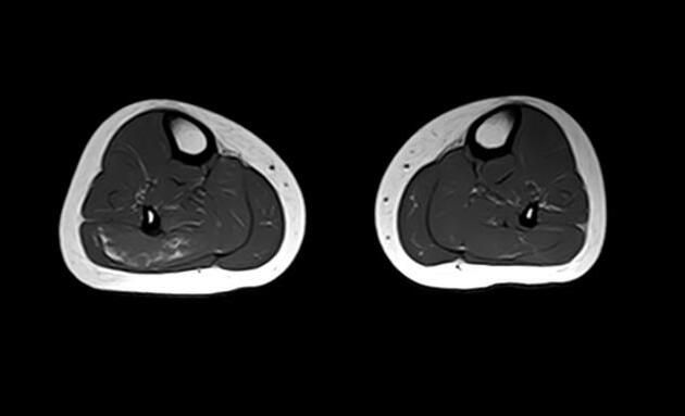
Well-circumscribed lobulated intramuscular soft tissue mass within the soleus muscle, isointense to muscles on T1, hyperintense on T2 and STIR, containing fluid-fluid levels of blood products as well as calcifications (Phleboliths) of low signal. The postcontrast sequences show progressive and heterogeneous enhancement. Normal thickness and signal intensity of the adjacent fibular cortex. Normal appearance of the surrounding muscles.
Case Discussion
MRI features most consistent with an intramuscular haemangioma.
Intramuscular haemangiomas, also known as intramuscular angiomas or intramuscular capillary-type haemangiomas, are rare intramuscular vascular lesions consisting of benign vascular channels within skeletal muscle. There is a peak in diagnosis in young adults and adolescents.