
Presentation
Painless swelling of the right parotid region. Ultrasound exam was inconclusive.
Patient Data
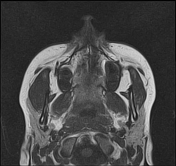

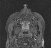
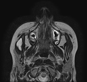
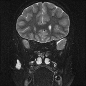
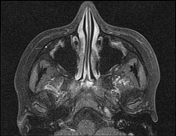
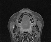
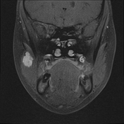
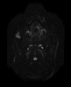
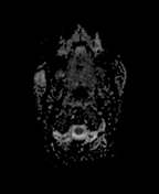

A small lobulated soft tissue mass is noted within the right masseter muscle, isointense to the muscle on T1WI, markedly hyperintense on T2WI with progressive enhancement and complete fill-in on delayed postcontrast sequences. No restricted diffusion is seen on DWI/ADC.
Case Discussion
MRI features characteristic of an intramuscular haemangioma within the masseter muscle.
Haemangioma within the masseter muscle may be confused with a parotid tumour or other muscular lesions. MRI exam is the modality of choice to precise its origin, characteristics features as well as its extent.
Additional contributor: Houssam-eddine Kouachi, Radiographer CIM Aurés, Batna, Algeria


 Unable to process the form. Check for errors and try again.