
Presentation
Renal mass on ultrasound.
Patient Data
Age: 85 years
Gender: Male
From the case:
Intraspinal dermoid cyst

Download
Info
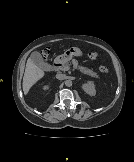
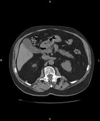
4.5 cm left lower pole renal mass.
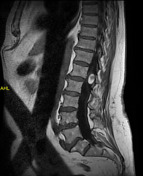
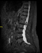
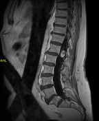
Non-enhancing mixed attenuation lesion in the spinal canal at the level of T12.
From the case:
Intraspinal dermoid cyst
Download
Info
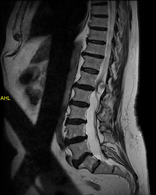
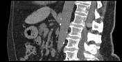
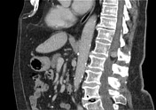
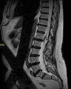
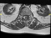
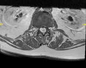
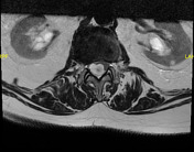
2.5 cm intradural, extramedullary mass dividing the filum terminale just distal to the conus. Fat and soft tissue composition. No enhancement.
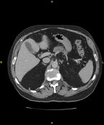
4.5 cm left lower pole renal tumour.
Case Discussion
The appearances are in keeping with an incidental spinal dermoid cyst identified at the time of assessing for a renal mass (a renal cell carcinoma).
This is one of the less commonly observed intradural, extramedullary masses.


 Unable to process the form. Check for errors and try again.