
Presentation
Was admitted to the emergency hospital for abdominal pain and dark red bloody stools, 2-3 times a day. Blood tests upon admission showed hemoglobin (HGB) of 95 g/L. No bleeding lesion was found colonoscopy and fibrogastroduodenoscopy.
Patient Data


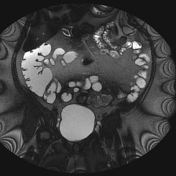
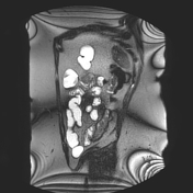
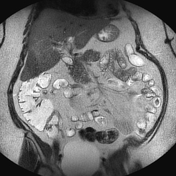
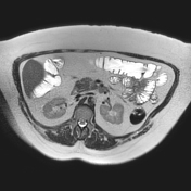
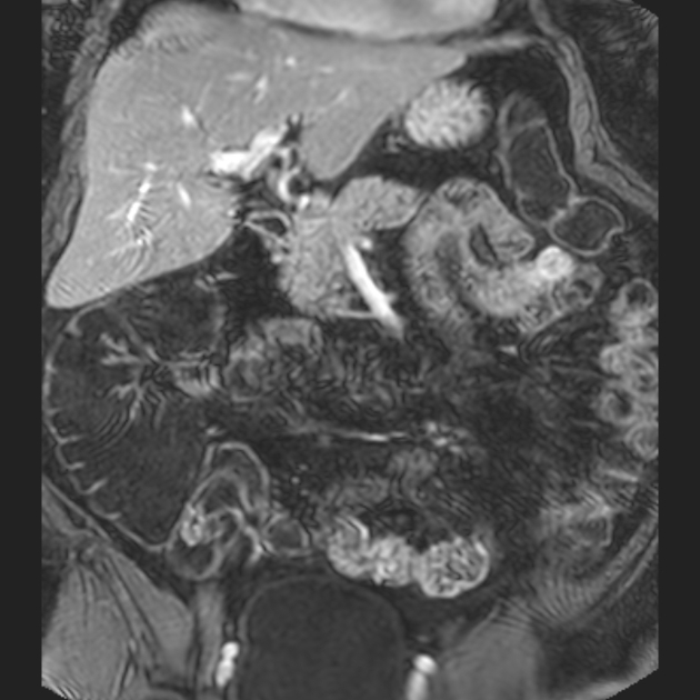
MR enterography with oral contrast using polyethylene glycol.
Mass in the jejunum, measuring 4 x 5 x 3 cm. Most of the mass lies on the side of the serous membrane, a smaller part protrudes into the lumen of the intestine. The appearances are highly suggestive of a gastrointestinal stromal tumor in the jejunum.
A wedge-shaped resection of the tumor-bearing jejunum was performed
Histology:
MICROSCOPIC DESCRIPTION: fusiform tumor of mesenchymal nature. The tumor grows into the mucous membrane of the jejunum. Moderate cellularity is noted. No necrosis. Low mitotic index (3 mitoses x 50 HPF).
In order to verify the tumor, immunohistochemistry was performed using antibodies CD117, CD34, SMA, which demonstrated overexpression CD117 (c-kit) and SMA in the cytoplasm of tumor cells, the absence of expression in tumor cells CD34 b S100.
Сonclusion: Morphological and immunophenotic features of the tumor define it as a stromal tumor of the jejunum/GIST. ICD-0 code 8936/2 (pT2).


 Unable to process the form. Check for errors and try again.