
Presentation
Acute change in mental status. Evaluate for intracranial abnormality.
Patient Data


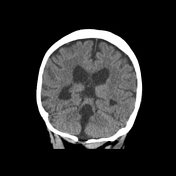

Elongation of the superior cerebellar peduncles, enlargement of the fourth ventricle and absent cerebellar vermis. The lateral and third ventricles are unremarkable.
The prominent CSF space along the cerebral convexities bilaterally is unchanged and could reflect cystic hygromas.
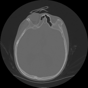
Posterior calvarial defect. There is a shrunken and partially calcified left globe compatible with phthisis bulbi. There is moderate paranasal sinus mucosal thickening.
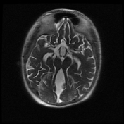

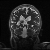

Thickening and elongation of the superior cerebellar peduncles in the axial plane, extending perpendicular to the brainstem resulting in the so-called “molar tooth” sign. Severe vermian hypoplasia with a cleft of CSF separating the cerebellar hemispheres which are formed. Enlarged fourth ventricle with a batwing configuration.
Supratentorial brain shows moderate ventriculomegaly at the lateral and third ventricles compatible with longstanding hydrocephalus.
Status post repair of occipital encephalocele with complete surgical closure.
Case Discussion
This is a case of Joubert syndrome with the classic "molar tooth" appearance of the midbrain, seen on both CT and MR.


 Unable to process the form. Check for errors and try again.