Kfuri and Schatzker classification of tibial plateau fractures
![](https://prod-images-static.radiopaedia.org/images/59914625/00d6e86924f9953c7dde5aeb79306d0bcd2a6616c5fde6b45a0122ed2b716b30_thumb.jpeg)
![](https://prod-images-static.radiopaedia.org/images/59914626/897a597587b12d2927b9e5a0b64c2fb85365d02ef8c5169d4958a99f6b7e587c_thumb.jpeg)
![](https://prod-images-static.radiopaedia.org/images/59914625/00d6e86924f9953c7dde5aeb79306d0bcd2a6616c5fde6b45a0122ed2b716b30_big_gallery.jpeg)
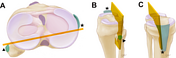
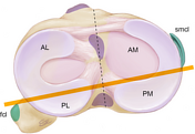
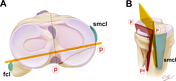
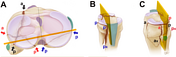
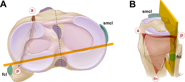
The virtual equator is defined by a plane intersecting the insertion of the fibular collateral ligament (fcl) on the lateral tubercle of the fibular head and the posterior aspect of the insertion of the superficial medial collateral ligament (smcl).
The virtual equator divides the medial and lateral axial columns into four quadrants.
![](https://prod-images-static.radiopaedia.org/images/59914627/8c121b1d2df690dc86bb7ecfb33a14a549451f684c13bccb8051922a0cdc9f70_thumb.jpeg)
![](https://prod-images-static.radiopaedia.org/images/59914628/db11fa7bc83962328d685c5a7554c897f8a5fd4a24ee50ec5c4693387529918b_thumb.jpeg)
![](https://prod-images-static.radiopaedia.org/images/59914629/a9885c20c06d2c9c72d21175965554e383cf73189abf7f94a501e4806311cdf9_thumb.jpeg)
![](https://prod-images-static.radiopaedia.org/images/59914630/7ae7ea6a449b52ae9142aa0733d9acce3d76db418fa413214dc728052cefefc4_thumb.jpeg)
![](https://prod-images-static.radiopaedia.org/images/59914631/e4533375711dc27de50ea12c0801ca53ad63b46fe19f4ad1e5ebfffbac69fb0a_thumb.jpeg)
![](https://prod-images-static.radiopaedia.org/images/59914632/9368c721df2ec7c0181cce28bd81418eff10b22eb7115c32b2a70294db89cb86_thumb.jpeg)
![](https://prod-images-static.radiopaedia.org/images/59914627/8c121b1d2df690dc86bb7ecfb33a14a549451f684c13bccb8051922a0cdc9f70_big_gallery.jpeg)
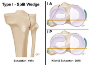
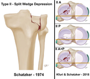
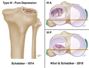
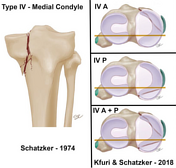
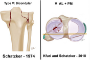
Illustrations of the six types of the original Schatzker classification with examples of the inclusion of the new modifiers.
![](https://prod-images-static.radiopaedia.org/images/59914633/fb9fc286d9c049cab3aa0226062ee998a347e94dc5755a105369a82f10b0943f_thumb.jpeg)
![](https://prod-images-static.radiopaedia.org/images/59914634/bc9bb585e27aaaecea1935e03572cf8a2b0f52acee7c1fdb0a9e6272ac4756c4_thumb.jpeg)
![](https://prod-images-static.radiopaedia.org/images/59914635/a7372778c53ec92c6bda84614e31826eed80130f5f71ca82c7799019c67ef212_thumb.jpeg)
![](https://prod-images-static.radiopaedia.org/images/59914636/3a066a31de888ab7b0194a4de611e75cc5d5121474195f919a9481cd6c7a3d34_thumb.jpeg)
![](https://prod-images-static.radiopaedia.org/images/59914633/fb9fc286d9c049cab3aa0226062ee998a347e94dc5755a105369a82f10b0943f_big_gallery.jpeg)
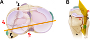
Examples of the Kfuri and Schatzker classification and the concept of the main wedge(s) plane(s).
The lowercase red letters mark the vertices of the wedge plane. These points where the wedge bisects the articular rim of the tibial plateau are marked as "a" if anterior to the virtual equator, and as "p" if posterior to it. The third point where the fracture exits at the metaphyseal area is denoted as "x", which could be anterior (ax) or posterior (px). These three points determine the main fracture plane.
Author: Stacy Turpin Cheavens.
Source: Kfuri M & Schatzker J. Revisiting the Schatzker Classification of Tibial Plateau Fractures. Injury. 2018;49(12):2252-63. https://doi.org/10.1016/j.injury.2018.11.010
Licence: CC BY-NC-ND 4.0
Case Discussion
The Kfuri and Schatzker classification is a revision of the classic Schatzker classification on tibial plateau fractures.
It complements the classic radiographic classification with the tridimensional interpretation of the fracture using CT.
For further details on the classification please read the reference article: Kfuri and Schatzker classification of tibial plateau fractures.