
Presentation
Seizures, swallowing difficulties, low cognitive performance, motor deficit, and gait worsening.
Patient Data

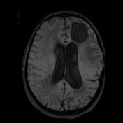
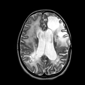
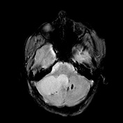
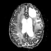

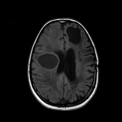
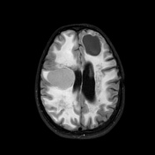
There are extensive abnormal white matter signal changes, indicating leukoencephalopathy, which is noted in the bilateral cerebellar, basal ganglia, as well as in both cerebral hemispheric white matter.
There are numerous small asymmetric calcifications scattered in the cerebellum, basal ganglia, and white matter of all lobes of the brain, which are hypointense and show blooming on SWI.
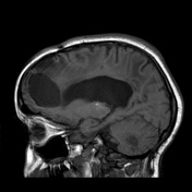
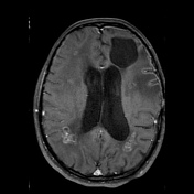
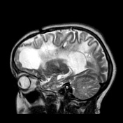
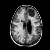
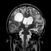
The two most significant cysts are at the right cerebellar hemisphere and the left frontal lobe, which are hypointense on T1W and hyperintense on T2W, without diffusion restriction. These cysts do not promote mass effect. There are small heterogeneous cystic lesions with surrounding oedema, and some of them associated with eccentrical calcifications. After the administration of contrast medium, there is multiple ring-like enhancement along the cyst walls and a few areas of focal enhancement in the degenerated white matter.
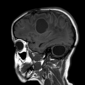
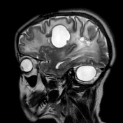
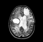
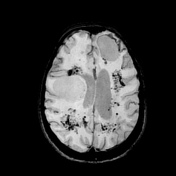
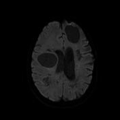
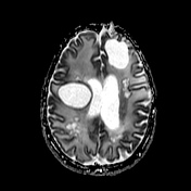

There is progression of the lesions, with persistent widespread white matter signal changes, further enlargement of the extensive coarse calcifications, and some cysts have enlarged gradually in size .
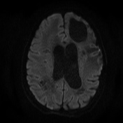
The cyst located in the right hemisphere demonstrates a higher signal than CSF on FLAIR, reflecting high protein content; it causes compression to the adjacent structures such as the right lateral ventricle, as well as effacement of local sulci. It was tiny on the last magnetic resonance study.
The diffusion-weighted image shows no water restriction in the cysts and affected white matter, and ADC demonstrates higher diffusivity, refecting tissue damage compatible with increased water content.
Case Discussion
Labrune syndrome is a rare progressive disease with unknown aetiology, which is characterised by a radiological triad of oedematous leukoencephalopathy, brain calcifications, and parenchymal cysts, detected by neuroimaging studies 1-6. There is an association with mutations in the SNORD118 gene 6.
The present case illustrates the features of Labrune syndrome, with a follow-up study demonstrating the evolutionary aspects of the appearance of the lesions.
Case courtesy
- Erick Cavalcante, MD - PGY-3, radiology resident, Department of Radiology
- Suzana M B Serra, MD – paediatric neurosurgeon, Department of Neurosurgery
- Mirela Gadelha, MD – Radiologist, Departament of Radiology
- Silvio Litvin, MD – Radiologist, Department of Radiology
- Antonio Rodrigues de Aguiar Neto, MD - Radiologist, Department of Radiology
Hospital da Restauração – Recife, PE – Brazil


 Unable to process the form. Check for errors and try again.