
Presentation
Left frontal swelling. No history of trauma or malignncy.
Patient Data
Age: 8 months
Gender: Male
From the case:
Langerhans cell histiocytosis


Download
Info

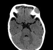
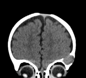
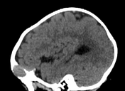
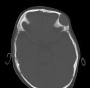
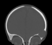
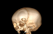
An expansile lesion is seen at the left frontal bone with soft tissue density and scalloped bony margins. No fat density or calcifications inside.
Case Discussion
A solitary well-demarcated lytic lesion involving both the inner and outer tables of the skull ... Considering the patient's age, eosinophilic granuloma is suggested as the first possibility, however intradiploic epidermoid cyst can be considered in the differential diagnosis.
MRI can help in the differentiation between them through the detection of diffusion restriction in the case of intradiploic epidermoid cyst.
Histopathology is better for a definite diagnosis.


 Unable to process the form. Check for errors and try again.