
Presentation
Puffy eyelid.
Patient Data
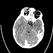

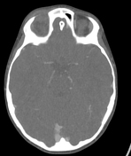
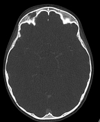
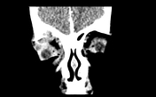
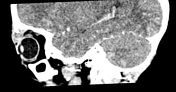

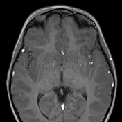
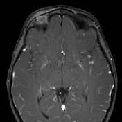
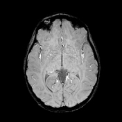
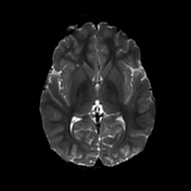
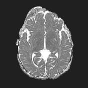
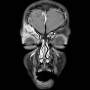
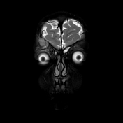
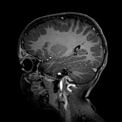
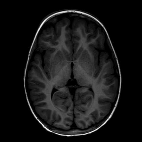
Lobular soft tissue mass measuring 1.3 x 1.5 x 2.5 cm at the superolateral aspect of the right orbit. The mass is heterogeneous, with cyst-like areas showing peripheral enhancement; displaces the ocular globe downward and slightly outward; involves the superior rectus muscle; does not involve the optic nerve; erodes bone at the lateral aspect of the orbital roof. Intracranial extension cannot confidently be ruled out.
Mucosal thickening in left frontal and maxillary sinuses and in several ethmoid cells and sphenoid sinus bilaterally.
The differential diagnosis in mainly of Langerhans cell histiocytosis (LCH) or, less likely, an aggressive focal infection.

Case Discussion
Came in because of a puffy eyelid for the past 3 weeks, not responding to treatment. On examination, a small mass was palpated. Sent for CT of the orbits.
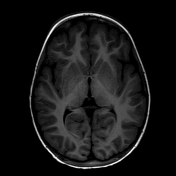
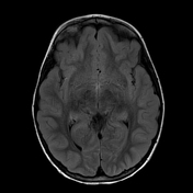
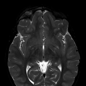
A right orbital mass was seen on CT and MRI.
Histopathology report:
Langerhans cell histiocytosis.
The tumour cells are strongly positive for CD1a and S100 and negative for CD68 (positive only within the multinuclear giant cells).
BRAF mutation not detected.
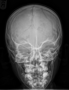
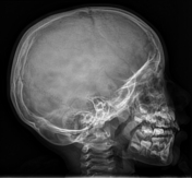
A skeletal survey was done, which showed no evidence of additional bone lesions. The skull X-rays are included here.


 Unable to process the form. Check for errors and try again.