
Presentation
Known case of retroviral disease. Headache since 2 months.
Patient Data
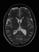


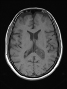
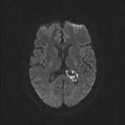
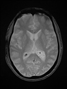
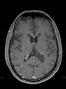
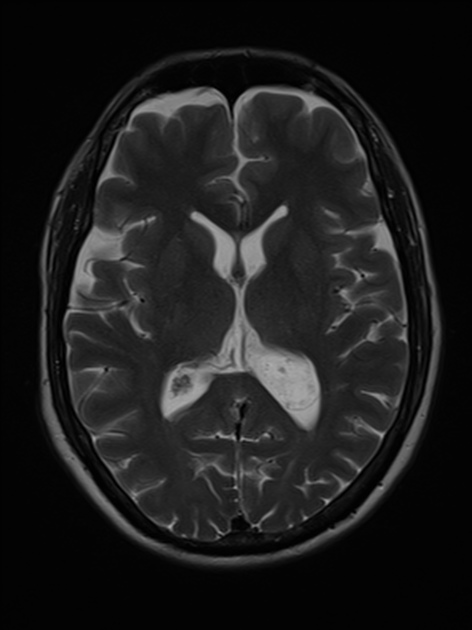
Plain and contrast MRI of brain show a well-defined T2 hyperintense, T1 hypointense and FLAIR heterogeneously hyperintense intraventricular lesion arising from the choroid plexus of left lateral ventricle measuring 3.1 x 1.8 x 2.0 cm (AP x TR x CC). There are scattered foci of restricted diffusion and blooming (calcifications) on gradient imaging. No periventricular CSF seepage is seen. There is no appreciable contrast enhancement on post-contrast T1 image.
These features are consistent with choroid plexus xanthogranuloma.
Case Discussion
Choroid plexus xanthogranulomas are benign lesions, usually located in lateral and third ventricles. Most of the lesions are asymptomatic and incidentally found. Third ventricular lesions can cause obstructive hydrocephalus.


 Unable to process the form. Check for errors and try again.