
Presentation
Palpitations.
Patient Data
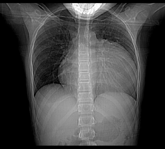
Significantly increased cardiothoracic ratio. Lung volumes and vascularity appear normal. The chest radiograph is suggestive of cardiomegaly versus pericardial effusion versus anterior mediastinal mass obscuring the left cardiac border.


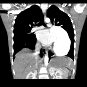
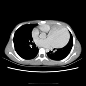
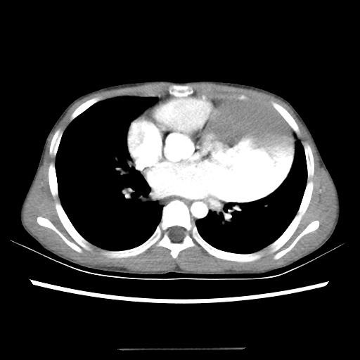
There is a large left atrial appendage aneurysm with swirling contrast inside. The aneurysm measures 9 x 7 x 13 cm (AP x T X CC) and diameter of its neck is 2 cm (nicely seen on coronal images).
The anterior aspect of the lesion shows homogeneous contrast filling on the portal phase, apart from a small area in the dependent anterior aspect, which represents a thrombus.
Case Discussion
The patient was diagnosed with a left atrial appendage (LAA) aneurysm. He was urgently referred to a tertiary hospital, where surgical resection was performed successfully.
LAA aneurysms are very rare; approximately 100 cases have been reported thus far 1. They are usually diagnosed incidentally on imaging but do predispose to atrial tachyarrhythmias and thromboembolism. Definitive treatment consists of surgical excision 2 or clipping 3.
Take-home message:
The mnemonic for the differential diagnosis of anterior mediastinal mass - 5Ts:
- Thymic lesions
- Thyroid lesions
- Teratoma
- Terrible lymphoma
- Thoracic aneurysms, which can be of aortic or cardiac origin


 Unable to process the form. Check for errors and try again.