
Presentation
Cough, fever, elevated WBC count, and chest pain.
Patient Data
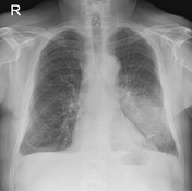

PA chest x-ray shows a large area of consolidation in the left lower zone. The heart border is sharp, the left hilum border is blurred and part of the left diaphragm is obscured.
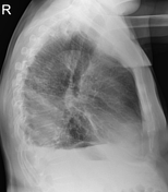
Lateral chest x-ray shows increased attenuation due to consolidation in the lower posterior part of the lung (spine sign).
Case Discussion
Typical x-ray features of left lower lobe pneumonia (with the typical clinical scenario).
This case demonstrates that even a PA x-ray and the utilization of a silhouette sign can localize lung pathology in 3D.
The PA projection alone is sufficient to localize pneumonia:
left lung, basal and posterior
the heart border is crisp (no silhouette sign), thus the consolidation is not in contact with the heart, therefore the pneumonia is in the dorsal part of the lung
If pneumonia is in the anterior basal part of the lung, the heart would be blurred.


 Unable to process the form. Check for errors and try again.