
Left-sided IVC with azygos continuation and retroaortic right renal vein
Presentation
Inguinal swelling. Past history of right inguinal hernia repair.
Patient Data


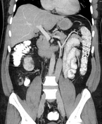
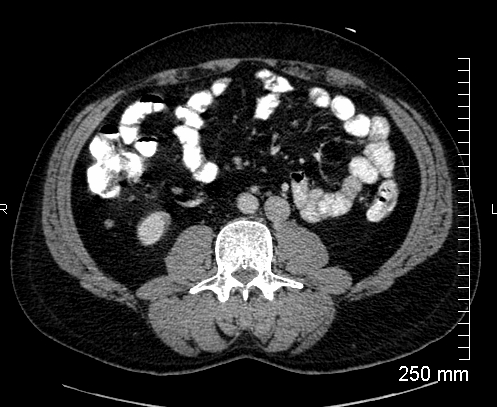
Recurrent right inguinal hernia containing fat with evidence of previous surgery is noted. Fat containing left inguinal and umbilical hernias. Small benign looking left adrenal nodule, likely an adenoma. Infra-renal IVC on the left side of the abdominal aorta. Right renal vein crosses the midline behind the aorta to drain in to the left-sided IVC. Left IVC crosses the midline behind the aorta above the renal arteries and continues as the azygos vein that enters the posterior mediastinum along with the aorta. Supra-hepatic IVC is formed only by the hepatic veins and has a usual drainage into the right atrium.
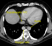
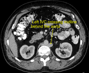
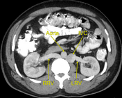

IVC=inferior vena cava, RRV=right renal vein, LRV=left renal vein.
Case Discussion
Left-sided IVC has a prevalence of 0.2–0.5%. It usually ends at left renal vein and crosses midline anterior to the aorta to join the normal pre-hepatic segment of IVC. This case shows left IVC with retro-aortic right renal vein, which has a prevalence of up to 2.1% as an isolated abnormality. Retro-aortic course of the renal vein can cause compression of the renal vein between the spine and aorta leading to the so called “posterior nutcracker syndrome”1.


 Unable to process the form. Check for errors and try again.