
Presentation
Abdominal distension and discomfort.
Patient Data
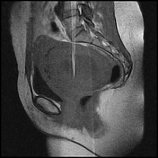

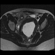
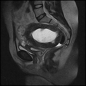
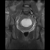
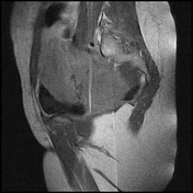
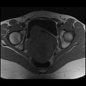
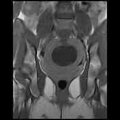
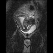
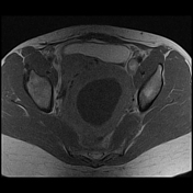
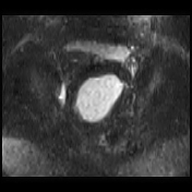
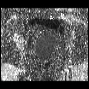

A large thick-walled left adnexal mass lesion is seen measuring about 11.7 x 10 x 13 cm along maximal AP, TS & CC dimensions, it shows a cystic centre that elicits bright T2, low T1 signal and thick peripheral wall that shows heterogeneous post-contrast enhancement, a pedicle is seen connecting the lesion to the posterior uterine wall. No evidence of aggressive features. No enlarged pelvic lymph nodes.
Case Discussion
When the leiomyoma gets increased in size, the vascular supply to its centre decreases, then it can undergo degeneration, which includes different types such as cystic, hyaline, red, and myxoid degenerations. Cystic degeneration is the least common type while hyaline degeneration is the commonest type of degeneration.


 Unable to process the form. Check for errors and try again.