
Presentation
Fell from scaffolding one hour ago resulting in pain and restricted motion in the left wrist.
Patient Data
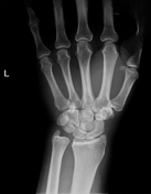

Abnormal radiocarpal arcs. On PA image projection, the lunate bone has an abnormal triangular appearance.
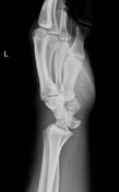
On lateral projection, the lunate bone is displaced ventrally, dislocated from the radius and capitate. Minimally displaced fracture of the styloid process of the radius.
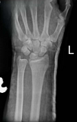
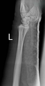

The lunate bone was reduced using an open surgical approach and a cast was applied.
The lunate bone has returned to its anatomical position.
Case Discussion
Lunate bone dislocation is a serious wrist injury. It is characterised by pain, swelling, deformity, and limited range of motion. This injury typically occurs after falls, sports injuries, and motor vehicle accidents.
The gold standard for diagnosis is x-ray examination. At this time on the lateral projection the lunate bone resembles an overturned cup, the spilt teacup sign. On the PA projection both perilunate and lunate dislocations cause rotation of the lunate bone which has a triangular appearance called the piece of pie sign. However, to determine specifically which type of dislocation is present, a lateral projection is necessary.
The injury requires immediate medical intervention to prevent complications such as nerve compression, wrist dysfunction, chronic pain and circulatory disorders.
Treatment involves either open or closed reduction.


 Unable to process the form. Check for errors and try again.