
Presentation
Gradually increasing abdominal distension after surgery for VP shunt placement.
Patient Data
Age: 10 years
Gender: Male
From the case:
Malfunctioned VP shunt


Show annotations
Download
Info
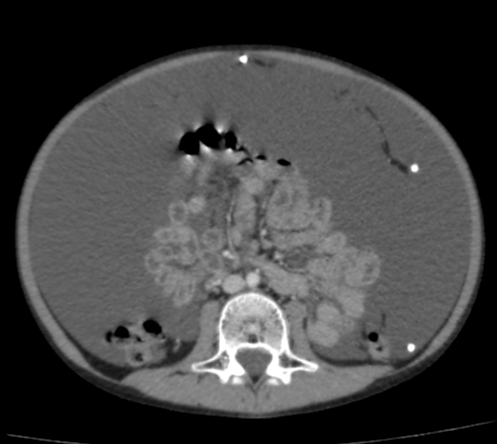
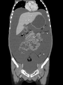
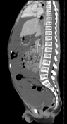
Gross abdominopelvic ascites is noted.
The peritoneum is thickened with significant enhancement, best visible in the upper abdomen.
No pseudocyst of abscess formation around the tip of the VP shunt (abdominal end) is seen.
Case Discussion
CT features in this case are of gross abdominopelvic ascites. However, no pseudocyst or collection formation around the tip of the VP shunt is seen.
The peritoneum is thickened and shows significant enhancement that suggests an infective process which is the most common cause of VP shunt malfunction in pediatrics.
Co-contributor: Dr. Anwar-ul-Haq Zadran.


 Unable to process the form. Check for errors and try again.