
Presentation
Shortness of breath. Attempted left internal jugular catheter placement.
Patient Data

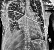
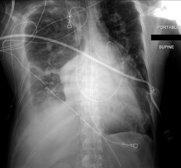
Endotracheal tube, right internal jugular catheter, and pulmonary artery catheter in place. The left internal Jugular catheter courses over the expected location of the aorta and the left pulmonary artery. Right pleural effusion, bibasilar consolidation. Enlarged heart.
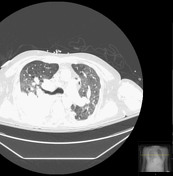

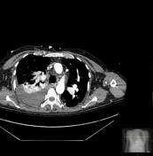
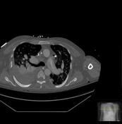
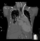

Left internal jugular catheter passes through the left internal jugular vein and courses extravascularly through the mediastinum along the aorta and pulmonary artery, with its tip just to the left of the main pulmonary artery. Small pneumomediastinum.
Large ventricular septal defect (VSD) with dilated left and right ventricles, right ventricular hypertrophy, and enlarged pulmonary arteries suggestive of pulmonary hypertension.
Endotracheal tube, right internal jugular catheter, and pulmonary artery catheter in place. Right pleural effusion, bibasilar consolidation.
Case Discussion
Central lines are, of course, supposed to be in either the SVC or at the cavoatrial junction. Placement through a vessel and into the mediastinum is rare. In this case, the catheter was removed without complication.


 Unable to process the form. Check for errors and try again.