
Presentation
Seizures. History of term normal delivery. Macrocephaly noted at six months of age. Started having seizures around 1 to 2 years of age, including behavioural arrest as well as secondary generalised tonic-clonic seizures. She also had a mild developmental delay. She was scanned at the time of the displayed study due to an increasing number of seizures that were becoming refractory to medications.
Patient Data
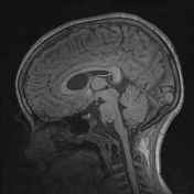

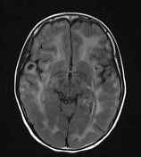
Confluent expansile T2 prolongation and FLAIR hyperintensity throughout the deep and subcortical white matter of both cerebral hemispheres, most prominent in the frontal, temporal and parietal lobes.
Additionally, there is T2 prolongation involving parts of the corpus callosum and posterior half of the posterior limb of the internal capsules bilaterally
Relative sparing of the fornices, optic radiations, anterior limb of the internal capsule and U-fibres in the parietal and occipital lobes (better appreciated on the fraction anisotropy maps).
Deep gray matter structures are also spared.
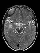
Multiple cysts are seen bilaterally in the subcortical white matter of the anteromedial aspect of the inferior temporal gyrus and lateral aspect of the superior temporal gyrus characterised by T2 hyperintensity which nulls on the FLAIR images, differentiating them from rest of the expansile white matter change.
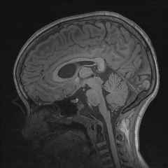
Mild T2 prolongation involving the white matter of the cerebellar hemispheres and adjacent to the fourth ventricle, without significant expansion as seen in the supratentorial white matter.
Patchy T2 hyperintensity in the cerebral peduncles and the white matter in the anterior two-thirds of the upper pons.
Associated global volume loss as demonstrated by prominent extra-axial spaces adjacent to the sulci as well as mildly enlarged ventricular system size.
Incidental cavum septi pellucidi and tiny T2 hyperintense/T1 hypointense 2 mm pars intermedia cyst.
Case Discussion
Findings are diagnostic of megalencephalic leukoencephalopathy with cysts (formerly called Van der Knapp leukoencephalopathy).


 Unable to process the form. Check for errors and try again.