
Presentation
Acute right hemiplegia, aphasia and confusion.
Patient Data
- Note: This case has been tagged as "legacy" as it no longer meets image preparation and/or other case publication guidelines.
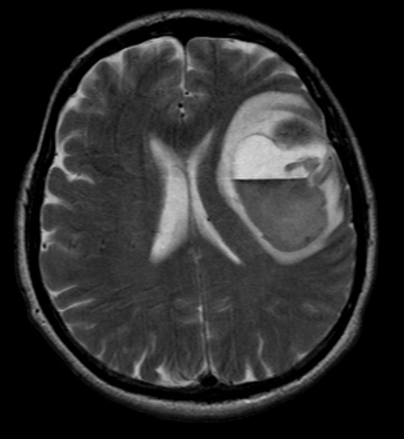
Large cerebral hematoma with a fluid-fluid level. There is a smaller low signal area anteriorly which corresponded to the known metastasis. This smaller area enhanced after gadolinium, as did the overlying dura.
Case Discussion
This patient has a known cerebral melanoma metastasis in the left frontal lobe.
Melanoma metastases may be hyperintense on T1W images and demonstrate signal loss on T2 or T2* sequences. This is due to the presence of both melanin and blood products. Melanoma metastases are 5x more likely to show signal loss on T2* images than lung metastases, and 4.5 times more likely to be T1 hyperintense. T1 hyperintensity correlates with melanin content better than does T2* signal loss. T2* imaging (or susceptibility-weighted imaging) may be useful in screening for melanoma metastases as lesions are more conspicuous.
Image contributed by: Dr Laughlin Dawes.


 Unable to process the form. Check for errors and try again.