
Presentation
Optic neuropathy.
Patient Data
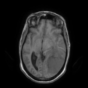

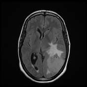
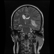
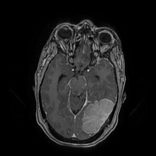
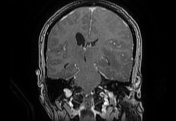
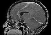
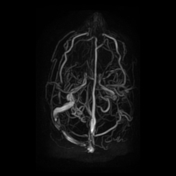
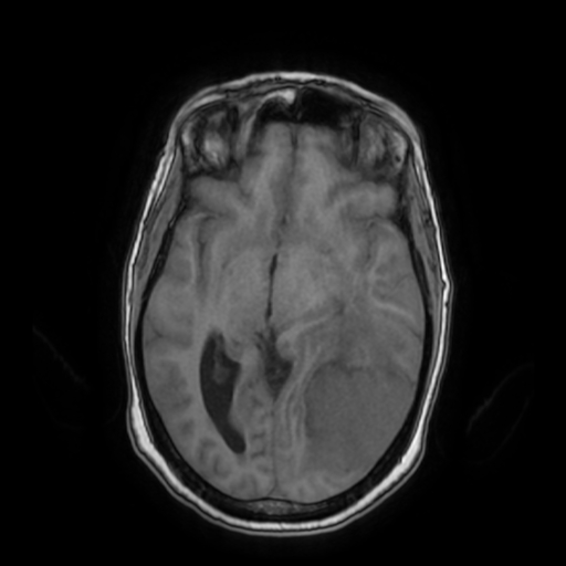
Large extra-axial mass centred on the left tentorium with a broad-based dural attachment. It demonstrates an iso-signal intensity to the grey matter on T1WI, high signal intensity on T2WI and FLAIR with relatively homogeneous enhancement with "dural tail sign" following IV contrast administration. Note mild hyperostosis with enhancement of adjacent bone. A thin rim of CSF "cleft sign" surrounding the lesion, indicating its extra-axial location. This mass invades the adjacent left transverse and sigmoid sinuses as well as the trocular herophili. A perilesional oedema is noted with mass effect on midline structures and dilatation of the contralateral ventricular system.
Case Discussion
MRI features consistent with a meningioma invading the adjacent dural venous sinuses


 Unable to process the form. Check for errors and try again.