
Presentation
History of chronic headaches. A CT scan was performed (not available) showing obstructive hydrocephalus with a pineal mass. A VP shunt was inserted and an MRI was requested for characterisation.
Patient Data


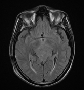
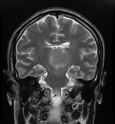
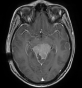
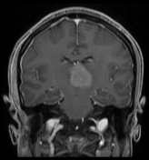
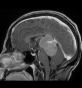
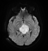
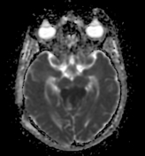
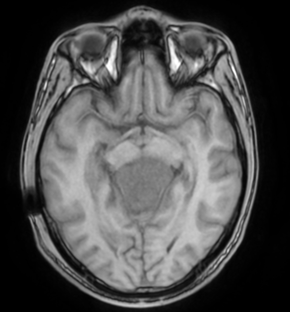
The MRI sequences demonstrate a large well-circumscribed lobulated mass centred on the pineal region measuring (5.5 x 5.3 x 4.3 cm). It elicits an isosignal to the cortical grey matter on T1/T2 and FLAIR vivid contrast enhancement following IV contrast and restricted diffusion on DWI/ADC. A mass effect is noted on the midbrain mainly the tectal plate with compression of the aqueduct of Sylvius. The vein of Galen/internal cerebral vein as well as the basal vein of Rosenthal are patent, coursing through the mass well-visualised on postcontrast sequences.
Case Discussion
MRI features of a pineal mass probably a meningioma.
On imaging, the pineal region masses have a relatively broad differential due to the presence of a variety of cell types found in this region (see pineal region masses).
Additional contributor: R. Bouguelaa, MD


 Unable to process the form. Check for errors and try again.