
Presentation
Worsening occipital headache for the past 3 weeks. No nausea or vomiting. No increased sensitivity to light. No fever or trauma. No previous history of migraines. Unremarkable neurological examination.
Patient Data
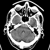

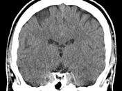
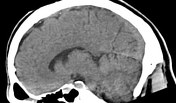
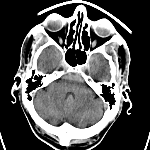
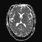
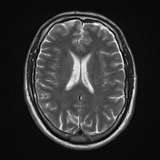
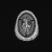
Normal intracranial appearances. No mass lesion, infarction, haemorrhage, hydrocephalus or extra-axial collection.

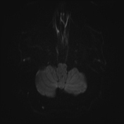
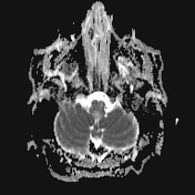
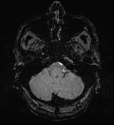
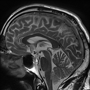
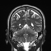
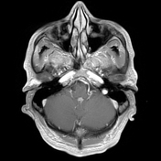
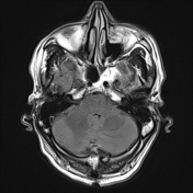
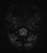
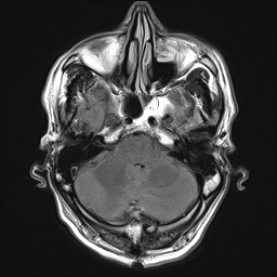
7x10x15mm (APxMLxCC) fourth-ventricle-mass, which is isointense to cortex and shows homogenous contrast enhancement. No calcifications or blood products seen. No hydrocephalus.
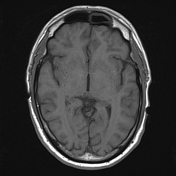
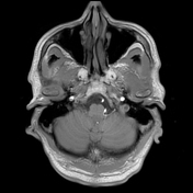
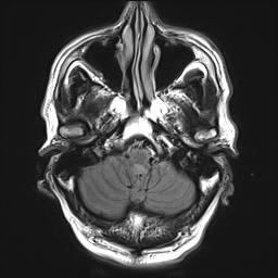
Postoperative MRI within 24 hours of the procedure:
Status post suboccipital craniotomy with related soft tissue changes.
Small amount of postoperative pneumocephalus is seen.
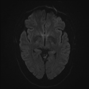
Minimal blood products in the fourth ventricle.
No acute ischaemia. No enhancing tumour rest. No hydrocephalus.
Case Discussion
The patient was given the option between surgical removal vs. reimaging in a few months, as the tumour was not thought of as the cause of the patient's symptoms. The patient chose the surgical removal of the tumour.
Immunohistochemistry expression profile:
- No IDH1 (R132H) expression
- Nuclear ATRX expression
Molecular-pathology expression profile:
- MGMT-promoter methylation
- Wildtype sequence of IDH1-Gene (R132) and IDH2-Gene (R172)
- Wildtype sequence of TERT-promoter (C228 and C250)
- No evidence of KIAA1549:BRAF fusion transcript Type A, B, C
Final Histological Diagnosis:
- Mixed Subependymoma/Ependymoma (WHO Grade 2)


 Unable to process the form. Check for errors and try again.