
Presentation
Headaches.
Patient Data


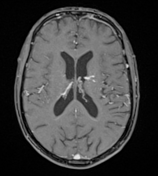
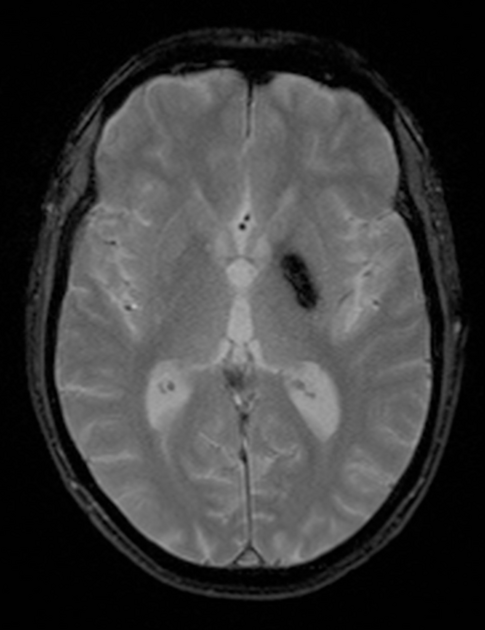
A well-defined oval-shaped mass is seen in the left pallidum, showing mixed signal intensity on T2, with the central high signal linned by a rim of hyposignal, along with prominent blooming on the GRE sequence. No evidence of surrounding oedema on FLAIR or enhancement after contrast administration.
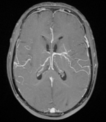
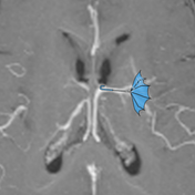
There is an adjacent vascular structure draining towards the frontal horn of the left lateral ventricle, with branches extending to the level of the ipsilateral insular cotex, leading to the characteristic caput medusae sign, palm tree appearance, or upside-down umbrella shape (best seen on the T1 C+ MIP sequence).


-
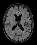
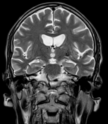
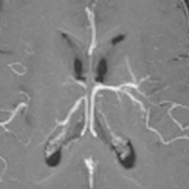
The images illustrate the different signs related to developmental venous anomalies:
image 2: palm tree appearance
image 3: inverted umbrella sign
Case Discussion
Incidental finding of a mixed vascular malformation.
Mixed vascular malformations are composed of a developmental venous anomaly and a cavernous malformation. Developmental venous anomaly is associated with cavernous malformation in 20% of cases. The main complication seems to be haemorrhage from the associated cavernoma.


 Unable to process the form. Check for errors and try again.