
Mobile CT brain post left posterior inferior cerebellar artery coiling following rupture
Presentation
Post aneurysm coiling. For mobile CT in the ICU.
Patient Data
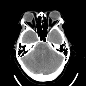

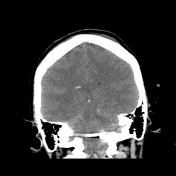

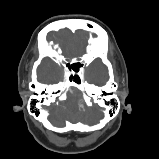
Post-procedure mobile CT brain in the intensive care unit demonstrating aneurysm coils in the region of the left posterior inferior cerebellar artery (PICA).
Extensive subarachnoid hemorrhage, which is predominantly within the subarachnoid cisterns and within both Sylvian fissures. Intraventricular extension of blood.
Right frontal approach EVD. No hydrocephalus.
Pre-procedural diagnostic CT circle of Willis angiogram demonstrating the ruptured left PICA aneurysm with extensive subarachnoid hemorrhage and intraventricular extension of blood.
Case Discussion
Mobile CT brains can be utilized effectively for gross pathology, particularly when patients are in intensive care and the time and human resources that are required to transport the patient to the radiology department are excessive.
This case nicely demonstrates adequate post-procedure imaging of the PICA aneurysm coils, the volume of blood as well as any mass effect or new hydrocephalus.


 Unable to process the form. Check for errors and try again.