
Presentation
Headache and transient ischaemic attacks since the age of 6 years.
Patient Data


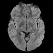
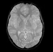
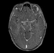
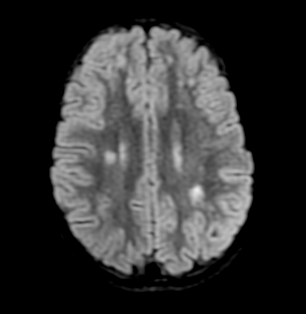
FLAIR images show bilateral multiple subcortical abnormal high signals located at the border zones between MCA and ACA as well as MCA and PCA and bilateral centrum semiovale, likely related to prior watershed ischaemic events.
No acute ischaemia or intracranial haemorrhage.
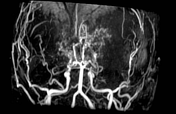
MR angiography of the brain shows:
occluded supraclinoid segments of both internal carotid arteries
stenosis of the proximal segments of PCAs
the circle of Willis and M1 segment of MCAs are replaced by a tortuous network of collaterals along their expected paths
extensive lenticulostriate, thalamostriate and cortical collaterals (puff of smoke sign)
no aneurysmal dilatation or arteriovenous malformation
Case Discussion
MRI findings are reflective of moyamoya disease.
The patient underwent surgery and had multiple skull burr holes (8 burr holes on each side) to allow the formation of local collaterals.


 Unable to process the form. Check for errors and try again.