
Presentation
Delayed mental and motor milestones. History of birth asphyxia.
Patient Data


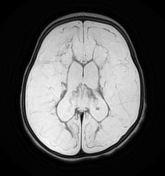
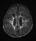
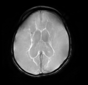
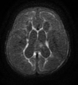
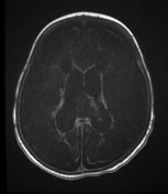
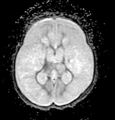
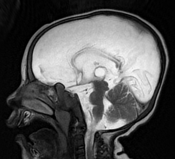
Both cerebral hemispheres parenchyma are replaced by multiple variable sized cystic areas of encephalomalacia involving the cortex and white matter.Ex vacuo dilatation of the lateral ventricles. Periventricular patches of gliosis eliciting high signal on T2 & FLAIR WI. Diffusely thinned corpus callosum. Preserved posterior fossa.
Opinion: Findings are impressive of multicystic encephalomalacia.
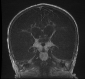
N.B: Enlarged nasopharyngeal adenoids.
Case Discussion
Multicystic encephalomalacia occurs in neonates after extensive brain insult resulting in loculated lacy pseudocysts within the white matter and cortex.
It is a common in neonatal hypoxic-ischaemic encephalopathy in a term neonate, including as a sequela of severe postnatal non-accidental head injury .


 Unable to process the form. Check for errors and try again.