
Presentation
Facial trauma, subsequent left orbital pain, and facial deformity with relatively decreased left eye visual acuity.
Patient Data
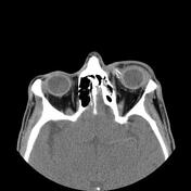

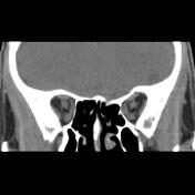
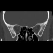
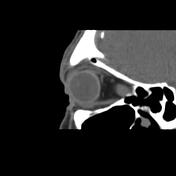

There are displaced multifocal and comminuted fractures in the left frontal calvarium inner and outer tables, orbital roof, frontoethmoidal sinus bones, medial orbital wall blow-out fracture extended within the ipsilateral ethmoid roof and optic canal, and related sphenoid sinus walls with the intraorbital and intracranial extension of displaced bones and impression on the corresponding left eye uveo-scleral layer and extraocular muscles.
Nasal deviation and proptosis of the left eye are seen.
Mild right eye nasal deviation is also seen.
The depressed fracture of the left side frontal calvarium with the depth of deformity up to 12 mm and impression on the frontal lobe is noted.
Soft tissue swelling of the left preseptal and lacrimal gland is seen.
Case Discussion
The case illustrates the non-contrast orbital MDCT features of concomitant frontal calvarium depressed fracture, orbital roof blow-in fracture, and orbital medial wall blow-out fracture, a relatively rare incidence. The case must be carefully evaluated from the point of intracranial haemorrhage.


 Unable to process the form. Check for errors and try again.