
Presentation
Recent headaches.
Patient Data


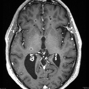
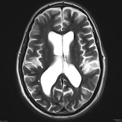
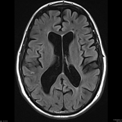
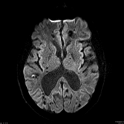
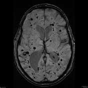
This MRI demonstrates widespread focal regions of susceptibility induced signal loss (best seen on susceptibility weighted imaging (SWI)) ranging in size from microscopic to a number of millimetres in diameter (remember that all T2* sensitive sequences result in a degree of blooming and thus grossly overestimate the actual size of the lesion).
One of the lesions in the right temporal lobe demonstrates high T1 and T2 signal centrally and perhaps a small amount of peripheral oedema, best seen on FLAIR. This suggests a recent bleed.
Case Discussion
This patient is from a known pedigree of autosomal dominant multiple cavernoma syndrome.
The differential for this case would include neurocysticercosis and cerebral amyloid angiopathy.
Against the diagnosis of neurocysticercosis is the variability in the size of the lesions, which by the time they calcify are typically more regular, measuring a few millimetres each. Furthermore there is no calcification in this case, although on the sequences provided that cannot be proven. Review of a CT (not shown) or phase images of the SWI (also not shown) would confirm this.
Cerebral amyloid angiopathy can have similar appearances however against this diagnosis is that A) the lesions, in this case, are distributed randomly throughout all parts of the brain, not having a predilection for the peripheral subcortical white matter which microhaemorrhage of cerebral amyloid angiopathy, and B) there is no evidence of prior larger lobar haemorrhage or superficial siderosis, both of which would be expected in this degree of change.
Case and discussion courtesy of Dr Frank Gaillard.


 Unable to process the form. Check for errors and try again.