
Presentation
Left foot swelling for the last 8 years. Non-tender, soft, mobile and stable in size.
Patient Data

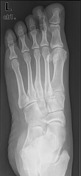
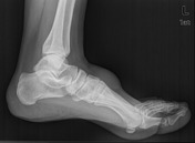
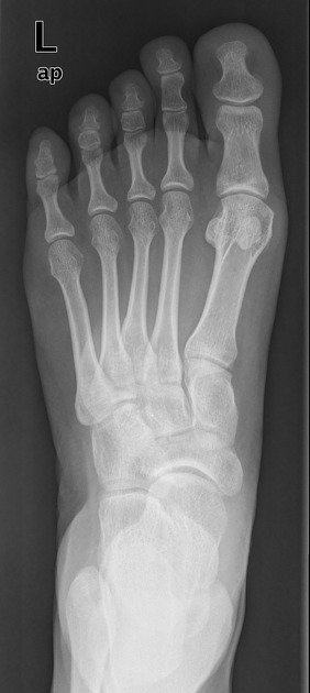
A well-defined non-calcified soft tissue swelling is seen along the dorsal aspect of the mid foot. No gross bony or articular abnormality is seen.


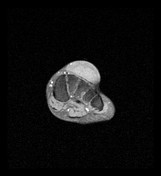
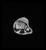
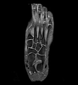
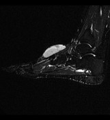
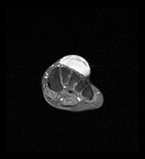
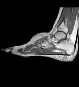
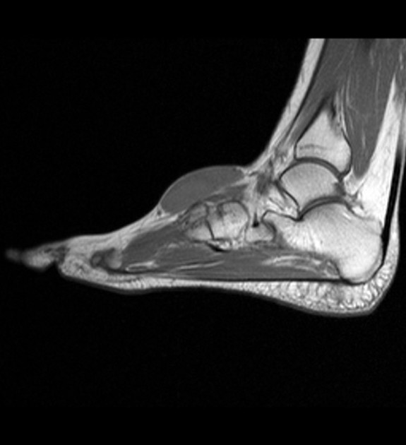
A fusiform subcutaneous soft tissue swelling measuring 5.5 x 1.8 x 4.2 cm is seen along the dorsal aspect of the foot at the level of tarsometatarsal. The lesion is isointense to the muscles on T1, markedly hyperintense on T2 fat-sat and slightly hyperintense on T2. The lesion shows good enhancement on post-contrast study and split fat sign to the related neurovascular bundle along the dorsal aspect of the foot raising the possibility of benign peripheral neurogenic tumour, like neurofibroma. No definite signs of invasion of nearby structures or scalloping of the underlying bone. The underlying extensor tendons are intact.
Case Discussion
Procedure: Excision.
Gross description: Specimen consists of a rounded mass of rubbery white tissue measuring 4.5 cm in maximum diameter. There is an overlying ellipse of skin 3.0 x 0.5 cm. Serial slicing shows a yellowish-white vaguely nodular cut surface.
Microscopy & immunohistochemistry: Cellular lesion composed of spindle cells with scanty cytoplasm and elongated & sometimes wavy nuclei. They are arranged in some areas in a storiform pattern. A few mitotic figures are seen and many blood vessels can be identified. A nerve is seen closely associated with the lesion. Immunohistochemistry shows spotty immunoreactivity to S100 and moderate diffuse immunoreactivity to NSE. There is also inexplicable positive reaction to CD34. The overall picture is in keeping with a benign peripheral nerve sheath tumour, neurofibroma.


 Unable to process the form. Check for errors and try again.