
Presentation
Left-sided headaches, nausea.
Patient Data
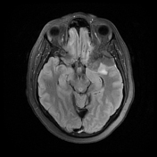

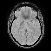
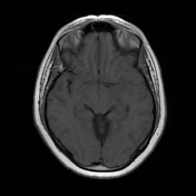
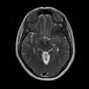

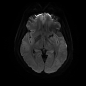
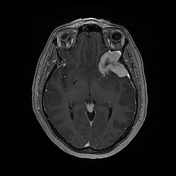
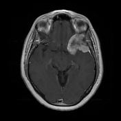

A T1-isointense and T2-hypointense enhancing dural-based, enhancing mass over the left sphenoid wing/middle cranial fossa measuring 4.3 x 4.5 x 5.0 cm. In the left aspect of the foramen magnum, there is a smaller 2.7 x 1.5 x 3 cm dural-based mass with similar signal characteristics and enhancement pattern.
Case Discussion
The patient underwent left pterional craniotomy for resection of the left sphenoid wing/middle cranial fossa dural-based mass. The final pathology demonstrated mass-forming sheets of epithelioid non-necrotizing granulomas, most consistent with sarcoidosis.
Upon further work-up, the patient did not have identifiable systemic sarcoidosis and was diagnosed with isolated neurosarcoidosis. Isolated neurosarcoidosis has been described in the literature as a rare occurrence (<1% of sarcoidosis cases).


 Unable to process the form. Check for errors and try again.