
Presentation
Dysphagia, decreased appetite, and weight loss for three months.
Patient Data
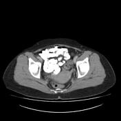

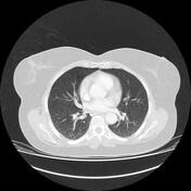
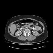
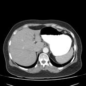
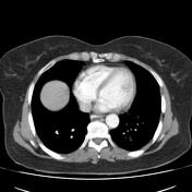
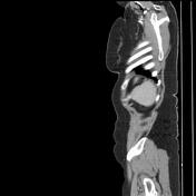
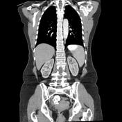
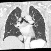
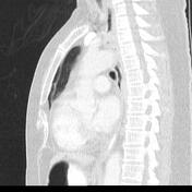
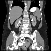
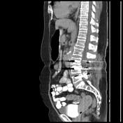

Solid mass lesion axial width up to 23 x 18 mm and height up to 32 mm in middle to the distal part of proximal thoracic esophagus with mild to the moderate enhancement and bulged within related mediastinal fat and with impression on adjacent trachea posterior wall and no obvious vascular invasion is seen. No obvious solid organ metastasis and no obvious mediastinal significant or suspicious lymph node are seen. Evidence of surgery on the distal segment of the lumbar spine due to a disc herniation is seen.
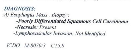
The biopsy result: poorly differentiated squamous cell carcinoma of the esophagus without lymphovascular invasion.
Case Discussion
The case illustrates the contrast-enhanced MDCT features of pathology-proved non-metastatic poorly differentiated squamous cell carcinoma of the esophagus.


 Unable to process the form. Check for errors and try again.