
Presentation
Acute abdominal pain and bloody stool.
Patient Data


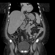
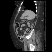
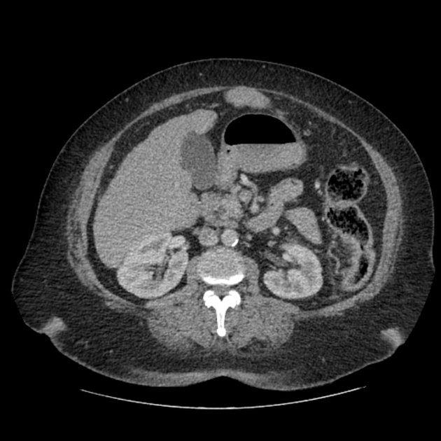
Colon wall thickening from distal transverse through proximal sigmoid with pericolonic fat stranding.
Patent superior and inferior mesenteric vessels. The inferior mesenteric artery is atherosclerotic and attenuated yet with no occlusion.
Case Discussion
The case shows the typical pattern of non-occlusive ischaemic colitis which starts from the splenic flexure through mid-sigmoid colon - a similar pattern to the IMA. Non-occlusive ischaemic colitis is likely secondary to transient hypoperfusion, and most commonly in the IMA distribution presenting with abdominal pain and blood in the stools. These typical imaging findings should be differentiated from "infection, inflammation" colitis or occlusive ischaemic colitis.


 Unable to process the form. Check for errors and try again.