
Presentation
6-year history of behavioral changes, diminished judgment, and executive dysfunction. Concern for possible dementia.
Patient Data
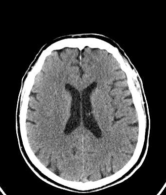
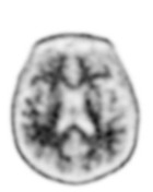
No acute findings on this head CT. No significant parenchymal atrophy. A few scattered hypoattenuating foci in the white matter, suggestive of microvascular changes.
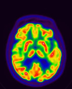

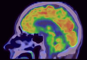
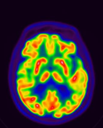
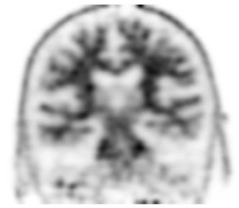
Physiologic radiotracer uptake throughout the brain parenchyma. Specifically, no areas of significantly reduced uptake suggestive of frontotemporal or Alzheimer's dementia.

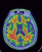
Normal radiotracer uptake through the brain parenchyma, with physiologic uptake outlining the white matter tracts. No abnormal areas of gray matter uptake are noted.
Case Discussion
Normal 18F-FDG and 18F-florbetapir PET/CT scans in this patient with concern for Alzheimer's or frontotemporal dementia.
Abnormal FDG scans demonstrate significantly decreased uptake in characteristic regions of the brain. For Alzheimer's dementia, the regions include the posterior parietal (precuneus) lobes, temporal lobes, and posterior cingulate gyrus. Later stages of the dementia demonstrate decreased uptake in the frontal lobes as well. For frontotemporal dementia, the regions include the frontal and/or temporal lobes (anterior greater than posterior), and anterior cingulate gyrus. Variants of these dementias and Lewy body dementia demonstrate decreased uptake in other characteristic areas (see references).
Abnormal 18F-florbetapir (Amyvid) or other amyloid PET radiotracer scans demonstrate increased uptake in the gray matter, suggestive of amyloid plaque deposition in these areas. A positive scan does not necessarily mean the patient has Alzheimer's dementia or that the patient will develop Alzheimer's dementia, it is simply a marker of plaque deposition. That said, it can help confirm clinical diagnosis of Alzheimer's dementia.
In this patient, there was a long-standing psychiatric history, which may have contributed to his presenting symptoms.


 Unable to process the form. Check for errors and try again.