
Presentation
Knee pain. Suspected chondral lesion.
Patient Data

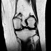
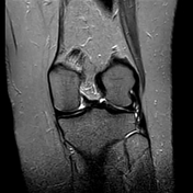
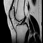
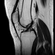


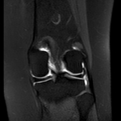
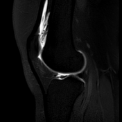
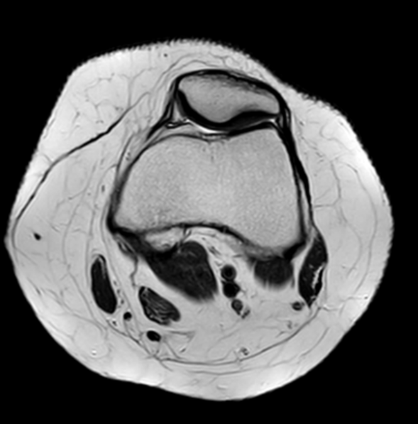
Degenerated posterior horn of the medial meniscus.
Otherwise, no meniscal or chondral lesions.
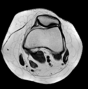
No intra-articular bodies. Intact cruciate and collateral ligaments. The arthrogram solution distends the knee joint.
Case Discussion
The patient was proposed a conventional MRI followed by an MR-arthrography with intra-articular injection of gadolinium.
MR-arthrography is better than conventional knee MRI, in meniscus and cartilage knee lesions.
The meniscal tears characterisation is better interpreted using MR-arthrography, especially in the detection of recurrent tears on operated menisci.
Also, it is helpful in the detection of cartilaginous lesions or foreign bodies in the joint space, and preoperative assessment before chondral repair procedures.


 Unable to process the form. Check for errors and try again.