
Presentation
Normal ankle MRI for reference
Patient Data
Age: 7 years
Gender: Female
From the case:
Normal pediatric ankle MRI

Download
Info

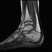
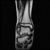
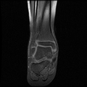
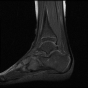
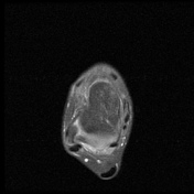
There is high PD fatsat signal intensity in the medial malleolus cartilaginous precursor.
There are multiple foci of normal red marrow rests in the talus and calcaneus.
There is a normal appearance of the physis. No joint effusion.
Case Discussion
Normal age features should not be confused with pathology.


 Unable to process the form. Check for errors and try again.