
Presentation
Dementia with gait disturbances.
Patient Data
Age: 80 years
Gender: Male
From the case:
Normal pressure hydrocephalus

Download
Info

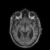
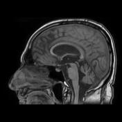
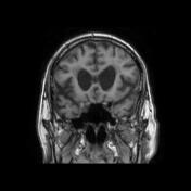
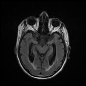
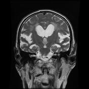
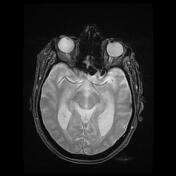
The MRI sequences demonstrate:
-
dilatation of the third and lateral ventricles with
upward bowing of the corpus callosum
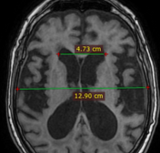
increased Evans index (0.36)
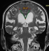
acute callosal angle (52°)
-
disproportionate enlargement of the subarachnoid space with
enlarged Sylvian fissures
narrow sulci and subarachnoid spaces at the vertex and parafalcine region
patchy FLAIR and T2 hyperintensities periventricular the white matter, subcortical region and centra semiovale suggestive chronic small vessel ischaemic change.
From the case:
Normal pressure hydrocephalus
Download
Info
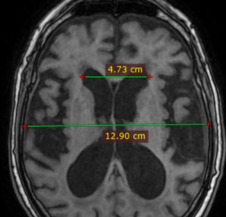
Annotated images:
Evans index = 0.36
callosal angle = 53°
Case Discussion
The clinical presentation and the MRI features are most consistent with a normal pressure hydrocephalus.


 Unable to process the form. Check for errors and try again.