
Presentation
The patient presented with a foreign body penetrating the left eye, accompanied by immediate pain and vision loss in the affected eye. There were no reported flashes or floaters. The left eye was erythematous, swollen, and bleeding upon arrival.
Patient Data
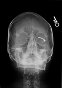
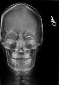
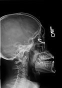

A curvilinear radiodense foreign body projects in the left orbit.
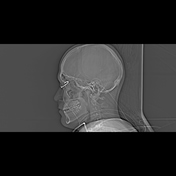
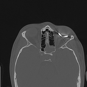

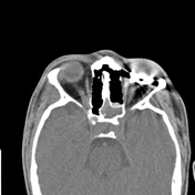
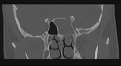
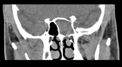
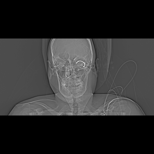
The nail enters the left orbit along the lateral margin of the globe and curves posteriorly and medially, terminating medial to the left globe. The nail disrupts the left posterosuperior globe, in the region of the retina. The adjacent extraocular muscles and the left optic nerve have a normal contour, with evaluation partially limited due to artifact from the nail. There is no intraconal fat stranding or retrobulbar hematoma.
No fracture is seen. Opacification of the left sphenoid sinus is likely chronic. There is a mucus retention cyst in the left maxillary sinus inferiorly.
Case Discussion
This case involves a 20-year-old male who presented with a penetrating injury to the left eye. The patient was using a nail gun on plywood when the nail ricocheted and penetrated the left globe.
The patient was taken to the operating room for left globe exploration and repair of the open globe injury/globe rupture. Preoperative ophthalmic evaluation revealed total retinal detachment. The nail was embedded in the sclera at the temporal aspect. The nail was carefully removed with mosquito forceps. The intraocular contents were reposited and a small amount of vitreous was excised due to hemorrhage. The sclera and conjunctiva were closed with sutures.
The postoperative course was uncomplicated, with pain effectively managed through oral medications. Antibiotics, particularly ciprofloxacin 500 mg twice daily for 14 days, were started to reduce the risk of infection.
Unfortunately, the patient lost all vision in the left eye at the time of the injury, and vision was never restored. A prephthisical globe was noted on follow-up exam. The patient declined enucleation, instead preferring a scleral shell.
Case co-authors: Yuliya Zayats and Emad Allam, Loyola University Medical Center


 Unable to process the form. Check for errors and try again.