
Presentation
Diabetic with peripheral neuropathy.
Patient Data
Age: 65 years
Gender: Male
From the case:
Osteomyelitis (interval imaging)

Download
Info
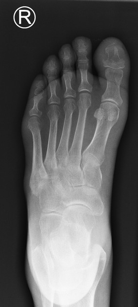
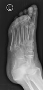
Lucency of the distal phalanx of the great toe with cortical destruction at the terminal tuft.
Minor soft tissue swelling of the distal great toe.
No vascular calcification.
{"current_user":null,"step_through_annotations":true,"access":{"can_edit":false,"can_download":true,"can_toggle_annotations":true,"can_feature":false,"can_examine_pipeline_reports":false,"can_pin":false},"extraPropsURL":"/studies/71655/annotated_viewer_json?c=1699857921\u0026embed_domain=hackmd.io%25252f%252540yipuafecsl2jsu8smr5njq%25252fbnjhjgjghjghjghfavicon.icofavicon.ico\u0026lang=gb"}
In the intervening 3 months, half of the distal phalanx of the great toe has been destroyed.
The soft tissue swelling remains.
Case Discussion
Diabetics with peripheral neuropathy are prone to osteomyelitis in the feet. This can range from minor phalangeal involvement right through to the necessity for amputation.
This interval imaging demonstrates how aggressive the bony destruction can be.


 Unable to process the form. Check for errors and try again.