
Presentation
Infertility.
Patient Data
Age: 35 years
Gender: Female
From the case:
Ovarian dermoid cyst - uterine adenomyosis with leiomyomas


Download
Info
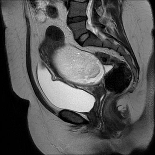
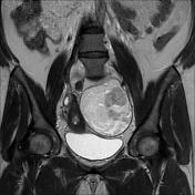
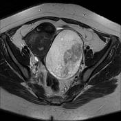
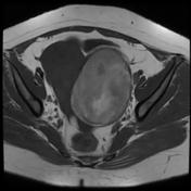
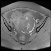
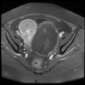
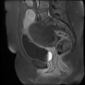
There is a large well-encapsulated ovoid mass arising from the left ovary, measuring 12 x 8 x 7 cm. It displays a complex signal on both T1 and T2, the main component is of fatty signal, attenuated on T1 fat sat with no enhancement on postcontrast sequences.
Thickening of the endometrial - myometrial junctional zone ranging from 14 to 20 mm with multiple uterine leiomyomas (Figo 4, 5 and 6).
Minimal effusion in Douglas pouch.
Case Discussion
The MRI features are most consistent with an ovarian dermoid cyst with uterine adenomyosis and leiomyomas.


 Unable to process the form. Check for errors and try again.