
Presentation
Pelvic pain.
Patient Data


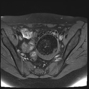
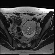
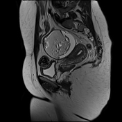
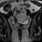
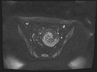
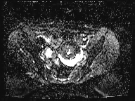
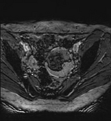
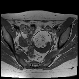
A large left ovarian cystic lesion with stretched ovarian parenchyma around its wall. It shows a predominate fat signal component (high signal intensity on T1 and T2 with complete suppression of signal on T1 fat sat sequence) and a fluid component (hypointense signal on T1 and high signal intensity on T2 and isointense signal on T1 fat sat sequence), with a fat-fluid level. No diffusion restriction identified. Areas of blooming are seen in SWI images.
Retroverted-retroflexed uterus with intrauterine contraceptive device.
Case Discussion
Ovarian mature cystic teratoma is the most common ovarian neoplasm in women in the childbearing period. It has a characteristic fat component that is confirmed on MRI by complete suppression of signal on fat-suppressed sequences. Blooming in SWI sequence may be due to the presence of calcifications.


 Unable to process the form. Check for errors and try again.