
Presentation
Abdominal lump with a pelvic mass on ultrasound. Tumor markers show elevated alpha-fetoprotein (AFP) and no beta-hCG secretion. MRI for further assessment.
Patient Data
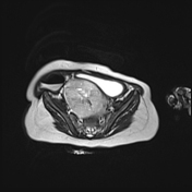

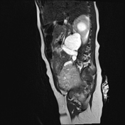

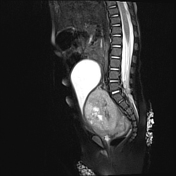
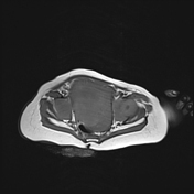
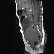
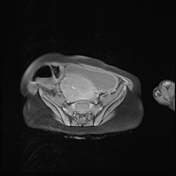
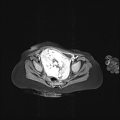
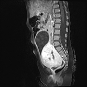
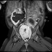

well-margined bulky pelvic solid mass, located between the urinary bladder and the rectum, arising either from the superior vaginal part or from the ovaries. This mass is T1 isointense (compared to muscle), and T2 hyperintense, measuring 62 x 54 mm on axial images and 61 mm on coronal images.
it demonstrates a vivid heterogeneous enhancement after Gadolinium injection.
marked ureteral and pelvicalyceal dilatation upstream of the pelvic mass
the uterus is present, of normal size considering the age, and looks unremarkable
there is no obvious rectal or urinary bladder extension
this mass is in close contact with the iliac vessels.
the ovaries are not seen
there is no lymphadenopathy nor ascites
Case Discussion
Features of a pelvic mass (of probable ovarian origin) with elevated AFP. In this case, the differentials include yolk sac tumor, ovarian embryonal carcinoma, and immature teratomas.
The patient was operated on and pathology confirmed an ovarian yolk sac tumor.


 Unable to process the form. Check for errors and try again.