
Presentation
Painful left parotid area swelling and fever since two weeks.
Patient Data
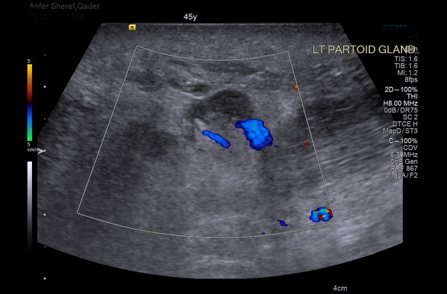
Diffuse skin and subcutaneous fat planes thickening with stranding as well as diffuse enlargement and edema of the left parotid gland. It has an ill-defined hypodense area related to the deep aspect of the superficial lobe. The lesion shows streaky irregular outlines and a central area of low density (collection) causing posterior enhancement. The lesion measures about 2.8 x 1.5 cm in diameter. Reactionary multiple enlarged cervical and submandibular lymph nodes are noted. The picture of diffuse left-side parotitis with an area of low echogenicity as described likely represents parotid abscess formation.


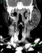
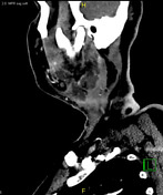
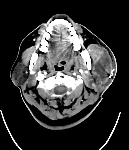
Three days later CT was done and a large cavitary lesion was seen affecting mainly the superficial lobe of the left parotid gland and reaching the deep lobe. It has heterogeneous peripheral enhancement. It involves and enlarges the left medial pterygoid and masseter muscles with posterolateral displacement of the left sternocleidomastoid muscle. The inflammatory lesion is effacing the left parapharyngeal fat without significantly compromising the related airway. The lesion enlarges and measures 6.4 X 5.7 X 9.4 cm in maximal dimensions with tiny air foci noted within. A radio-dense drainage tube is noted abutting the lateral surface of the lesion and noted in the related significantly thickened subcutaneous tissues. Subcutaneous fatty stranding with skin thickening is seen involving mainly the left aspect of the face and neck. Multiple enlarged bilateral cervical and submental lymph nodes, mainly on the left side at multiple levels.
Case Discussion
Enlarged left parotid gland with inflammatory changes harboring an intraglandular collection with marginal enhancement proved to be abscess formation.


 Unable to process the form. Check for errors and try again.