
Presentation
History of metastases of unknown primary. Prior PET-CT showed hepatic cysts versus hemangioma. For MRI assessment.
Patient Data
Age: 80 years
Gender: Female


Download
Info
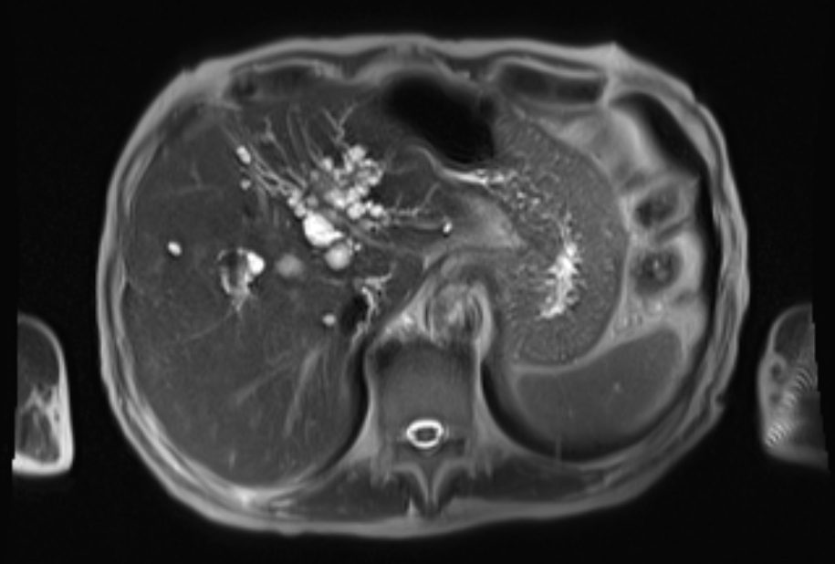
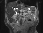
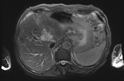
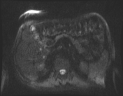
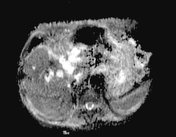
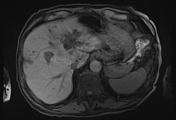
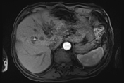
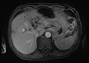
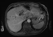
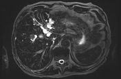
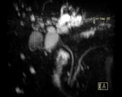
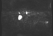
Clustered cysts are noted on both sides of the hilar portal vein and its left branch, largest reaching 1.5 cm. These are associated with:
- subtle biliary ectasia at its periphery
- small scattered cysts in all hepatic segments, all <1 cm
- no definite ductal communications
- no stones or masses
Case Discussion
Features of non-suspicious peribiliary cysts and small scattered simple hepatic cysts. Peribiliary cysts are a rare incidental finding of cyst formation around intrahepatic biliary ductules in a hilar distribution lacking any communication with the biliary tree.


 Unable to process the form. Check for errors and try again.