
Presentation
Chronic right ear discharge and diminished hearing. History of previous surgical intervention.
Patient Data
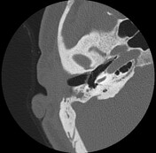

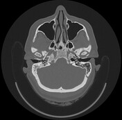
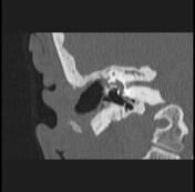
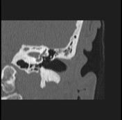

Previous operative right canal down mastoidectomy. Thickened tympanic membrane with circumferential soft tissue density was noted in the surgical bed and in the attic. Non-visualized ossicles. A linear hyperdense structure is seen reaching the oval window that should be correlated with the intra-op details. Abnormal bone formation seen within the attic is suggestive of tympanosclerosis. The deficient bony wall of the lateral semicircular canal suggests a labyrinthine fistula. Narrow facial canal (tympanic segment) with probable involvement in the labyrinthine fistula; otherwise normal morphology of the inner ear structures. Opacified mastoid air cells with thickened sclerosed trabeculae. The right external auditory canal shows minimal thickening of its lining, underlying bones are intact suggestive of inflammatory changes.
Case Discussion
A perilymphatic fistula (also known as a labyrinthine fistula) is an abnormal communication between the fluid-filled space of the inner ear and the air-filled space of the middle ear.


 Unable to process the form. Check for errors and try again.