
Presentation
Presents with 4 weeks of increasing abdominal distention, epigastric pain, and nausea. Self-reports history of hepatic cysts diagnosed in childhood. Denies recent weight loss, fevers, or night sweats. Elevated serum CA-125 levels noted.
Patient Data


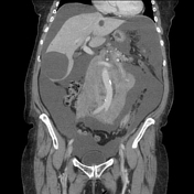
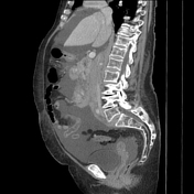
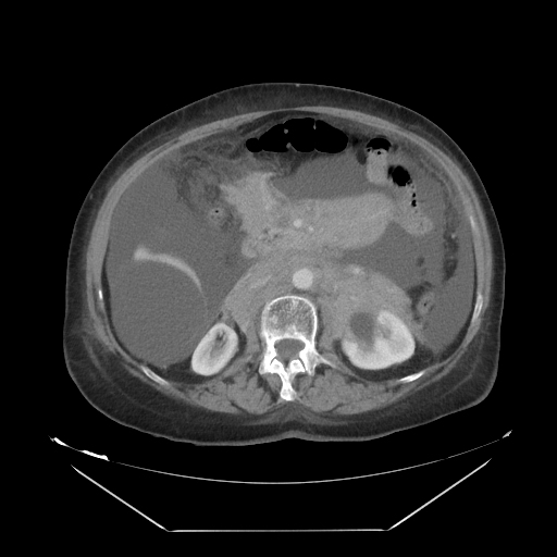
CT of the abdomen and pelvis with IV contrast demonstrates an irregular, bulky soft tissue mass involving the retroperitoneum and peritoneum. It encases the SMA, aorta, and IVC, and extends anteriorly to invade the mesentery. There is large volume ascites in all four quadrants, scalloping of the hepatic margins, and compression of the IVC. There is a sub-centimeter nodule, most likely subpleural in nature. Hepatic cysts are visible, largest in the inferior right hepatic lobe.
Case Discussion
Hospital Course:
Serum CA-125 was greater than 1500 U/mL, concerning for possible ovarian cancer. Serum CEA, CA 19-9, and beta-hCG levels were within normal limits.
Therapeutic paracentesis yielded 6.5 L of chylous fluid. Cytology was unremarkable. The patient then underwent a diagnostic laparoscopy.
Pathology:
MICROSCOPIC EXAM: The atypical lymphocytes are B cells strongly positive for CD20, CD79a, PAX5, CD10, BCL2, and weakly positive for BCL6. Ki-67 positivity is less than 10%. Stains are negative for cyclin D1, MUM-1, and cytokeratin AE1/AE3.
FINAL DIAGNOSIS: Classic follicular lymphoma, low grade
Discussion:
Low-grade follicular lymphomas are typically indolent in course, are usually advanced at the time of diagnosis, and are infrequently associated with 'B-symptoms' 1. Due to the high frequency of large-volume ascites at presentation, CA-125 elevations are not uncommon, which can lead to diagnostic confusion especially with advanced ovarian carcinoma 2. On imaging, advanced follicular lymphomas typically present as large, bulky masses which are typically homogenous in appearance and non-obstructive in nature, often encasing mesenteric vessels to produce a classic 'sandwich sign' 3. Additional findings commonly include involvement or invasion of the omentum and mesentery, splenomegaly, and extensive lymphadenopathy not necessarily contained to the peritoneum 4.
Timely tissue biopsy and careful assessment of radiologic findings is vital, as diagnostic confusion may result in unnecessary surgeries and delayed initiation of combination chemotherapy and rituximab. Appropriate treatment has been observed to drastically decrease tumor burden even in advanced cases 5.


 Unable to process the form. Check for errors and try again.