
Presentation
Ultrasound revealed an enlarged prostate. Blood tests showed elevated PSA levels (15.62ng/mL).
Patient Data
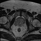

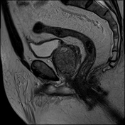
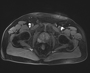

The prostate is enlarged, measuring approximately 51 x 54 x 42 mm (width x height x anteroposterior), with an estimated volume of ~60 ml. PSA density 0.26 ng/ml.
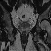
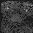
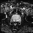
In the right posterolateral peripheral zone at the midportion level, there is a poorly defined nodule with low signal intensity on T2W imaging (≤14 mm in diameter), high signal intensity on DWI (b = 1400), and marked hypointensity on ADC (indicating true diffusion restriction). The nodule demonstrates early contrast enhancement on dynamic T1FS +C imaging.
Normal seminal vesicles. No pelvic nodes. No bone lesions.
Case Discussion
Imaging findings are consistent with a PI-RADS 4 lesion in the peripheral zone of the prostate. The patient subsequently underwent a biopsy, and histopathological results confirmed prostate adenocarcinoma.


 Unable to process the form. Check for errors and try again.