
Presentation
Left knee joint pain and swelling for one year.
Patient Data
Age: 20 years
Gender: Female
From the case:
Pigmented villonodular synovitis (PVNS)


Show annotations
Download
Info
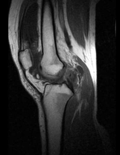
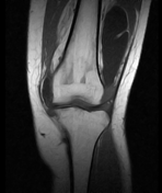
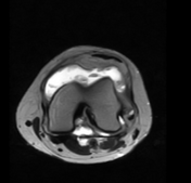
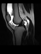
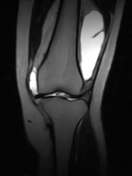
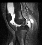
The synovium is showing irregular nodular thickening with evidence of dark areas suggesting hemosiderin deposition.
Moderate joint effusion is evident, more marked in the supra patella recess.
A lobulated fluid signals intensity area noted in the posteromedial aspect popliteal fossa with a narrow neck outline by gastrocnemius and semimembranosus tendons suggestive of Baker cyst. Fat planes with neurovascular bundles appear preserved.
Case Discussion
MRI features are most likely suggestive of pigmented villonodular synovitis along with Baker’s cyst and joint effusion as described above.
Co-contributor: Dr. Anwar-ul-Haq Zadran.


 Unable to process the form. Check for errors and try again.